
CALCIFICATION ON THUMB BASE ( OSTHEOARTHRITIS OF CMC JOINT OF THE THUMB)

1. CALCIFICATION ON THUMB BASE

(Osteoarthritis of the Thumb Basal Joint)

The thumb allows very special movements in a harmony with the other fingers. Most of these movements particularly pinching and gripping  are possible with a functional basal joint (carpometacarpal joint) of the thumb.  Because of its unique design it tends to develop earlier arthritis than those in the fingers. Destortion of this joint results in pain when it moves. Arthritis of thumb basal joint is seen more in women, older than 4o years of age.

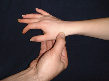
During physical examination, pain and loss of power whilst pinching or gripping, swelling and/or tenderness at the base of the thumb(picture 1), limitations in joint movements are noticed. Patient and doctor may feel friction if the thumb is tried to move in different directions when proksimal part is keeping stabil.

(figure 1)

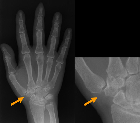
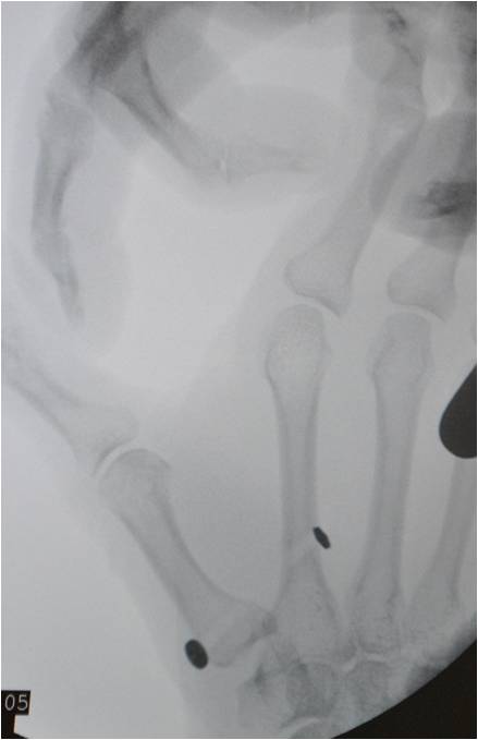
X Ray reveals distortion in congruence(dislocation) and cartilage surfaces of the joint, bony spurs(picture 2).

(figure 2)

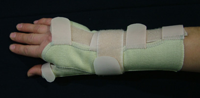
In the early stages, positive response could be obtained with non-surgical methods. Ice application several times a day, 5-15 minutes each time, aspirin or non-steroid antiinflammatory drugs may be helpful for pain and swelling relief. Supporting splints limit the joint movements and helps lessening the pain(picture 3). Steroid injections may be also helpful for pain relief.

(figure 3)

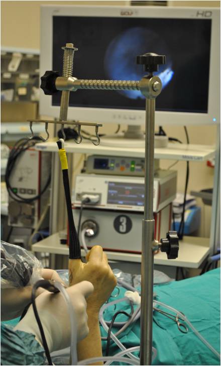
If  conservative methods fail, arthroscope assisted closed surgical approach is next treatment of choice(picture 4). The joint can be visualized directly by an arthroscopic camera. Very fine(1.9-2.4 mm diameter), high technology cameras and instruments are used for this surgery. Bony spurs can be shaved and hypertrophied synovia can be removed. Dislocation of the joint is corrected using a tight rope. Using this rope, first metacarpal is brought to its proper anatomical position with the carpal bone trapezium(picture 5). After surgery, no splint needs to be worn, and patient is allowed to use his or her hand as soon as possible. So, these patients can return to their work earlier.One of the main advantage of this surgery is to keep the commonly preferred methods as described below for the failed and severe cases.

For end stage cases (severe degenerations on the joint and bone surfaces) or elder patients whose bone dansity is poor, to remove the damaged parts or trapezium(carpal bone)is the solution. Some tendons are used for suspension of the thumb and to fill the empty space after bone removal. After this surgery, plaster splints are worn for several weeks. This surgery also works very well for pain relief. It only affects the grip power after surgery compared with the camera assisted method.

  • (picture 4)

  • (picture 5)

Continue reading

VASCULARİZED BONE TRANSPLANTATİON

Our patients suffer from bone loss due to some reasons. Bone loss is sometimes due to loss of bone content (osteonecrosis) due to the deterioration of the circulation of the bone, sometimes due to defects that occur due to non-union of the broken bone after the fracture (pseudoarthrosis or nonunion), sometimes as a result of the treatment of bone tumors with tumors removed by us. It may occur as a result of the defects created by the bone, and sometimes as a result of the defects created by us as a result of the removal of bone inflammation (osteomyelitis).

The bone tissue to be taken from the patient can be obtained from different body parts with or without vessels, depending on the size and characteristics of the defect to be applied. For example, for a defect formed as a result of removing a benign tumor from the finger bones, a 1 cm incision from the elbow region is sufficient, while for larger bone defects. vascular bone tissue from different body parts will be needed.

In vascular bone transplantation, the bone tissue taken is transported to the defective area with its vein. This means that the transferred bone is living bone. This process can be done by removing a bone section with its vein from a region close to the defective area and turning it into the defective area, or the bone taken from a region far from the defective area with its vein. It can also be done by suturing a vein in the area of ​​the defect under a microscope.

There are many advantages of carrying the bone alive with its vein. In order for the treatment to be successful in bone transplants, it is expected that the transferred bone will fuse to the applied area. Vascular (live) bone transplantation increases the chance of success of this union, that is, the treatment, especially in long bone defects larger than 8 cm, vascular bone transplantation It is very important for the success of the treatment.

A vascular tissue reduces the risk of infection as it allows the body defense cells and the antibiotics we give to the patient to reach this area. This situation increases the success of the treatment, especially in bone transplants for the treatment of bone infection (osteomyelitis).

Another common area of ​​use for vascular bone transplant surgeries is avascular necrosis (loss of bone) in which bone circulation is impaired. This may occur in small bones in the wrist (kienbock-lunatum avascular necrosis) or in the hip (femoral head avascular necrosis). Because it causes vascular bone transplantation to the damaged area, it is the most appropriate treatment for patients who can often be applied for the cause of the disease.

Although vascular bone transplants give very satisfactory results in suitable patients, a team with advanced microsurgery experience is required to perform these surgeries.

POLLICISATION

Pollicisation is the process of forming a thumb from the other fingers of the hand to replace the missing thumb. It is mostly applied because of loss due to trauma or congenital absence. Among the congenital anomalies, the most frequently pollicized condition is the developmental deficiency of the thumb (thumb hypoplasia) and the absence (or instability) of the joint between the metacarpal and the wrist bone (trapezium) below it. Apart from this, the giant thumb (macrodactyly), Congenital multi-finger and Mirror hand deformity are other congenital anomalies in which pollicisation treatment may be applied. After the pollicisation surgery, cortical plasticity, that is, the acceptance of the new finger as a thumb by the brain will be possible with the sprouting branches that will be sent by neurons (nerve cells) in the brain from the brain regions adjacent to the area where the thumb is represented. 1-1.5 years of age is the most accurate timing for pollicisation surgery in congenital anomalies and, as soon as possible after loss in traumatic cases. Thus, it will be possible to both experience the deficiency for the least amount of time and learn how to use of the new thumb.

The index finger is mostly used for pollicisation. Rarely, if both the thumb and index finger are absent and the current situation does not allow toe transfer to hand, then the middle finger can be pollicized as well. The finger to be used for transfer must not be affected by the existing anomaly or trauma. The transfer is performed by preserving all the structures of the finger to be pollicised. Preservation of arteries and veins is critical for the vitality of this finger. Sensory nerves, flexor and extensor tendons of the finger should also be properly protected in order to function after transfer. In normal hand structure, the thumb consists of two and the other fingers consist of three phalanges. In order for the transfered finger to adapt both visually and functionally, the metatarsal bone of the transfered finger is shortened appropriately and bone fixation is achieved in this way. The fixation of the bone, which is shaped by shortening and rotated a little, is made by means of surgical wires, one part of which is left out of the tissue. When it is seen that the reshaped bone is fused in the X-rays taken at the end of 4-6 weeks, the wires are removed during the dressing with a simple procedure.

Physiotherapy, which comes to the agenda after the completion of the surgical and post-surgical wound care process, has a critical importance as it is after most operations of hand surgery. The use of the new thumb as much as possible should be supported by play therapy methods in children and rehabilitation techniques in adults. With the increase in the use of the new thumb, the adaptation of the brain increases, as the brain adaptation increases, the patient can use his hand for finer Works more.

The most common complications after pollicisation are; The development of contracture between the new thumb and 2nd finger, the inadequacy of the tip-to-pulp touch of the thumb and the other fingers, which we call opposition, and rotation defects (malrotation) in the thumb. However, most of these problems can be overcome by secondary surgeries.

The thumb is responsible for about 40% of hand functions. Therefore, if it is not properly remedied in case of its deficiency or loss, it will result in a serious inability to use of the hand. Since the aim here is to provide a functional well-being rather than improving the appearance, pollicisation is one of the most accurate approaches in cases of loss and birth problems mentioned above.